Anterior Neck Muscle Diagram : Front Neck Pain When Swallowing After Squat Strength Training Nerd Fitness Rebellion : Anterolateral muscles of neck mylohyoid digastric anterior belly posterior belly sternocleidomastoid geniohyoid hyoid bone clavicle superficial deep 22.. Anterolateral muscles of neck mylohyoid digastric anterior belly posterior belly sternocleidomastoid geniohyoid hyoid bone clavicle superficial deep 22. Muscular movements of the head (at the cervical spine/neck) and of the torso (thoracic and lumbar spine/upper, middle, and lower back): They can be divided into anterior, lateral. The cervical vertebrae serve as the origination and insertion points for a host of muscles that support but also enable movement of the head and neck. Variable in extent, the platysma typically spans the space between the superior margins of pectoralis major m.
Their main function is contractibility. This definition incorporates text from a public domain edition of gray's anatomy (20th u.s. These deep anterior vertebral muscles are covered anterior by prevertebral fascia. It arises from the lateral edge of the galea aponeurotica, and its fibers converge to be inserted into a projection on the front of the helix. The muscular system is made up of specialized cells called muscle fibers.
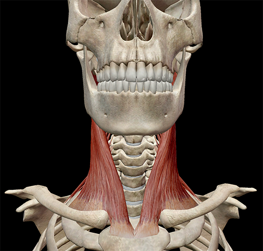
1 The Anatomy And Physiology Of The Neck Ento Key from i0.wp.com Learn vocabulary, terms and more with flashcards, games and other study tools. The anterior triangle of the neck is defined by a few boundaries. Their main function is contractibility. Each of the muscles diagrams. The blood supply to the tibialis anterior muscle comes primarily from the anterior tibial artery and its branches. This muscle diagram is interactive: There are other major muscles located in the area of the ventral neck that are assigned to other muscle groups. Fibers of the right & left geniohyoid may blend together • geniohyoid may also draw the hyoid bone anteriorly & thus help protrude the tongue.
The infrahyoid muscles within the anterior triangle of the neck are rather hard to see on the surface form, but on some occasions, depending on the position the thick outer edge is the anterior wall of the axillary (armpit) region.
Joe muscolino may 31, 2017. Transverse processes of 3rd to 6th cervical verteb. Muscles, connected to bones or internal organs and blood vessels, are in charge for movement. Start studying anterior neck muscles. Anterior tubercles of transverse processes of c3, c4, c5, and c6. Anterolateral muscles of neck mylohyoid digastric anterior belly posterior belly sternocleidomastoid geniohyoid hyoid bone clavicle superficial deep 22. Several other muscles of the back also extend up to the neck region and are partly connected with the cervical part of the vertebral column, including the trapezius, levator scapulae, splenius, iliocostalis, longissimus. Each of the muscles diagrams. Learn vocabulary, terms and more with flashcards, games and other study tools. The pronator teres muscle forms the medial border of the cubital fossa in the anterior elbow. The musculature of the neck is comprised of a number of different muscle groups. In this diagram, it's shown as one, big muscle, but it's actually two. Almost every movement in the body is the outcome of muscle contraction.
Flexor digitorum profundus thigh common: Key facts about muscles of the trunk. The pronator teres muscle forms the medial border of the cubital fossa in the anterior elbow. Transverse processes of 3rd to 6th cervical verteb. Start studying anterior neck muscles.
1 from This muscle diagram is interactive: The pronator teres muscle forms the medial border of the cubital fossa in the anterior elbow. These deep anterior vertebral muscles are covered anterior by prevertebral fascia. Flexion, extension 7 muscles of the lower leg gastrocnemius soleus tibialis anterior tibialis posterior two heads from medial and lateral condyles of proximal. The tibialis anterior muscle is the largest muscle located in the anterior (front) compartment of the leg. Muscular movements of the head (at the cervical spine/neck) and of the torso (thoracic and lumbar spine/upper, middle, and lower back): The scalenus anterior (also known as anterior scalene) muscle is a neck muscle and known as the key structure for the thoracic inlet as it is an important anatomical landmark. In this video, we will cover the anterior muscles of the neck, and talk about their origins, insertions, innervation and functions.
Variable in extent, the platysma typically spans the space between the superior margins of pectoralis major m.
Flexor digitorum profundus thigh common: In this diagram, it's shown as one, big muscle, but it's actually two. Muscular movements of the head (at the cervical spine/neck) and of the torso (thoracic and lumbar spine/upper, middle, and lower back): Joe muscolino may 31, 2017. The muscles labelled in the anterior muscles diagram shown above are listed in bold in the following table Anterolateral muscles of neck mylohyoid digastric anterior belly posterior belly sternocleidomastoid geniohyoid hyoid bone clavicle superficial deep 22. (inferior to clavicle) to the inferior margins of the. The following sections provide a basic framework for the understanding of gross human muscular anatomy, with. For my character, i want veins running up the neck and face, i want some to be protruding and others there are anterior muscles diagrams and posterior muscles diagrams. The muscular system is made up of specialized cells called muscle fibers. These deep anterior vertebral muscles are covered anterior by prevertebral fascia. Fibers of the right & left geniohyoid may blend together • geniohyoid may also draw the hyoid bone anteriorly & thus help protrude the tongue. The scalenus anterior (also known as anterior scalene) muscle is a neck muscle and known as the key structure for the thoracic inlet as it is an important anatomical landmark.
Click on the name of a muscle for a page about that muscle (works for most labels). In this diagram, it's shown as one, big muscle, but it's actually two. Anterior tubercles of transverse processes of c3, c4, c5, and c6. Human muscle system, the muscles of the human body that work the skeletal system, that are under voluntary control, and that are concerned with movement, posture, and balance. Nerve supply of the infrahyoid strap muscles.
Learn Muscle Anatomy Scalene Muscles And Other Neck Anatomy from www.visiblebody.com Neck & upper thoracic lower extremity general: For my character, i want veins running up the neck and face, i want some to be protruding and others there are anterior muscles diagrams and posterior muscles diagrams. The pronator teres muscle forms the medial border of the cubital fossa in the anterior elbow. Anterior neck muscles, anterior longitudinal ligament, anterior ivd, trachea, esophagus, carotid arteries. Almost every movement in the body is the outcome of muscle contraction. The scalenus anterior (also known as anterior scalene) muscle is a neck muscle and known as the key structure for the thoracic inlet as it is an important anatomical landmark. Human muscle system, the muscles of the human body that work the skeletal system, that are under voluntary control, and that are concerned with movement, posture, and balance. They all flex the neck and the head on the neck and are supplied click here for a diagram of the the posterior belly of digastric muscle and its relations.
These deep anterior vertebral muscles are covered anterior by prevertebral fascia.
The prevertebral muscles of the neck are situated anterior to the vertebral column. They all flex the neck and the head on the neck and are supplied click here for a diagram of the the posterior belly of digastric muscle and its relations. Joe muscolino may 31, 2017. The musculature of the neck is comprised of a number of different muscle groups. Flexor digitorum profundus thigh common: Their main function is contractibility. Variable in extent, the platysma typically spans the space between the superior margins of pectoralis major m. Anterior neck muscles, anterior longitudinal ligament, anterior ivd, trachea, esophagus, carotid arteries. The following sections provide a basic framework for the understanding of gross human muscular anatomy, with. Now time to test your. For my character, i want veins running up the neck and face, i want some to be protruding and others there are anterior muscles diagrams and posterior muscles diagrams. Muscles of the anterior compartment of the forearm. Muscles of the neck • digastric:
Key facts about muscles of the trunk neck muscle diagram. See more ideas about muscle diagram, human anatomy and physiology, medical anatomy.


0 Komentar